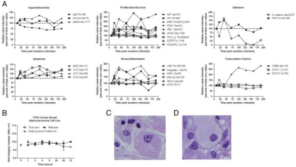
Figure 2.

A multi-purpose chemical solution for stabilization and preservation of proteins and RNA, and maintenance of histomorphology. (A) Frozen uterine leiomyoma tissue reveals on-going phosphoproteomic changes post excision. A reverse phase protein microarray analysis of tissue analyzed from a deep area of the tissue block (1.0–2.0 mm from surface) showed reactive protein changes as compared with the time zero sample (100% value, 10 min post excision, outermost tissue surface). Reactive proteins constituted a variety of molecular cascades: apoptotic pathway, stress/inflammation pathway, hypoxia/ischemia pathway, transcription factors, proliferation/survival pathway, and adhesion/cytoskeleton proteins. (B) RNA stability time course of T47D-cultured cells. T47D human mammary adenocarcinoma cell lines were cultured at 37°C, 5% CO2. Medium was removed and cells washed three times with cold phosphate-buffered saline. The cells were scraped from the flasks, combined and aliquots were incubated with RNAlater (Qiagen) (black square) or our multi-purpose fixative (black triangle) at 4°C for 1, 2, 4, 8, 24, 48, and 72 h. An untreated cell culture aliquot was used as the time zero control (open circle). RNA was extracted using the RNA Mini Kit (Qiagen) and RIN were determined using a Bioanalyzer 2100 (Agilent) for duplicate samples at each time point. (C and D) Tissue stabilized at room temperature in a multi-purpose chemical solution yields histomorphology similar to formalin fixed tissue. (C) Human breast tumor epithelium fixed in a multi-purpose stabilization solution containing phosphatase and kinase inhibitors, alcohol, and a permeation enhancer, was processed via a standard histology technique. H&E staining showed well-delineated nuclear membranes and chromatin clumping comparable to an adjacent tissue sample fixed in 5% formalin (D). (UltraLight Histology™ processing courtesy of Dr. Thomas Donndelinger, Bi-Biomics).
