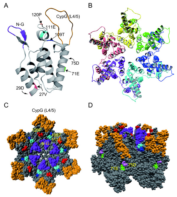
Figure 7.
Three-dimensional structural models of GH123 CA. (A) Structure of the N-terminal half of CA monomer. The model was constructed by homology-modeling using "MOE-Align" and "MOE-Homology" in the Molecular Operating Environment (MOE) as described previously [73,74]. N-G, dark purple; the 27thV and the 29thD, pink; Cyp G (L4/5), orange; the 71stE, green; the 75thD, light purple; the 109th T, dark blue; the 111th E, light blue; and the 120th P, red. The structure of CA hexamer from the top (B and C) and side (D) is shown.

