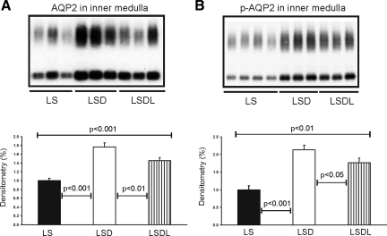
Fig. 2.
Semiquantitative immunoblotting of proteins prepared from the inner medulla. A: immunoblot is reacted with anti-aquaporin-2 (AQP2) and reveals 29- and 35- to 50-kDa AQP2 bands. Densitometric analysis reveals that the expression of inner medullary AQP2 is significantly increased in response to dDAVP treatment in the LSD group compared with control rats (LS), whereas AQP2 expression is significantly reduced in response to cotreatment of dDAVP and losartan in the LSDL group compared with LSD rats. B: immunoblot is reacted with anti-p-AQP2 (phosphorylated in the PKA-phosphorylation consensus site Ser-256) and reveals 29- and 35- to 50-kDa p-AQP2 bands. Densitometric analysis reveals that expression of inner medullary p-AQP2 is significantly increased in response to dDAVP treatment in the LSD group compared with LS rats, whereas p-AQP2 expression is significantly decreased in response to the cotreatment of dDAVP and losartan in the LSDL group compared with LSD rats.

