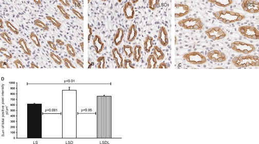
Fig. 3.
Immunoperoxidase microscopy of AQP2 in the inner medulla. AQP2 labeling is present at the apical and intracellular domains of the inner medullary collecting duct cells in LS (A), LSD (B), and LSDL rats (C). Strong apical immunoperoxidase labeling of AQP2 is observed in response to dDAVP treatment in the LSD group, whereas losartan cotreatment reduces the labeling of AQP2 in dDAVP-treated rats in the LSD group. Semiquantitative analysis of AQP2 immunoperoxidase labeling showed that the sum of total positive pixel intensity in LSD was significantly higher than that in LS, whereas losartan treatment decreased the intensity of AQP2 labeling in LSDL compared with LSD (D).

