Fig. 5.
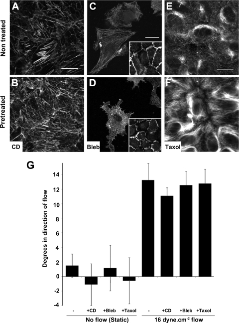
Flow-induced EC-cell junction inclination was independent of cytoskeletal structure. The cytoskeletal structure was disrupted by pretreatment of HUVEC confluent monolayers with 0.1 μM cytochalasin D (CD), 50 μM blebbistatin (Bleb), or 5 μM Taxol for 2 min, 30 min, and 2 h, respectively. Cytoskeleton disruption was verified by staining for phalloidin (A and B) and myosin IIA (C and D) and Taxol-induced inhibition of depolymerization of microtubules by acetylated-tubulin (E and F). Scale bars are 20 μm. Differences in myosin IIA staining were more detectable when using a lower cell seeding density where cytoplasmic retraction was observed (D). However, no cell retraction was observed on confluent monolayers. A wider PECAM-1 membrane staining including increased number of protusions after blebbistatin pretreatment was noticed (insets). G: steady shear stress of 30 min at 16 dyn/cm2 did not show any significant difference in EC-cell inclination after cytoskeletal disruption.

