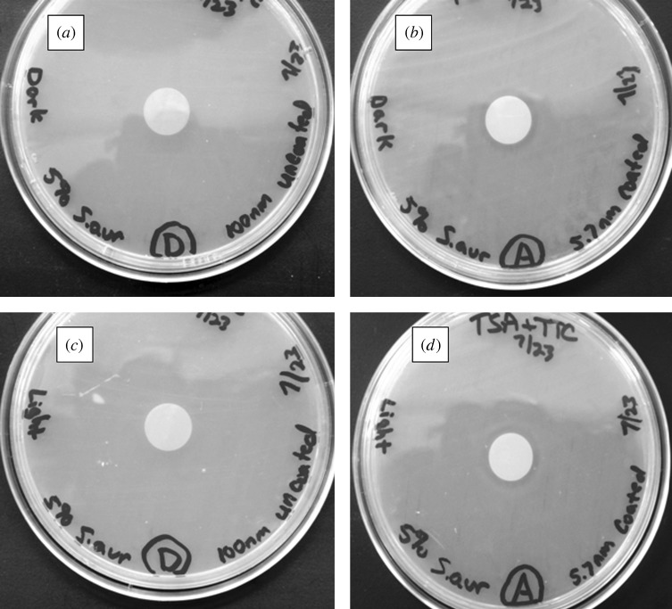
Figure 16.
Light microscopy images of agar plating assay results after 24 h of incubation. Materials were examined on tryptic soy agar plates, which were inoculated with S. aureus. (a) Uncoated 100 nm pore size nanoporous alumina membrane without light exposure. (b) Zinc oxide-coated (coating= 5 nm) 100 nm pore size nanoporous alumina membrane without light exposure. (c) Uncoated 100 nm pore size nanoporous alumina membrane under continuous light exposure. (d) Zinc oxide-coated (coating= 5 nm) 100 nm pore size nanoporous alumina membrane under continuous light exposure.

