
Figure 5.
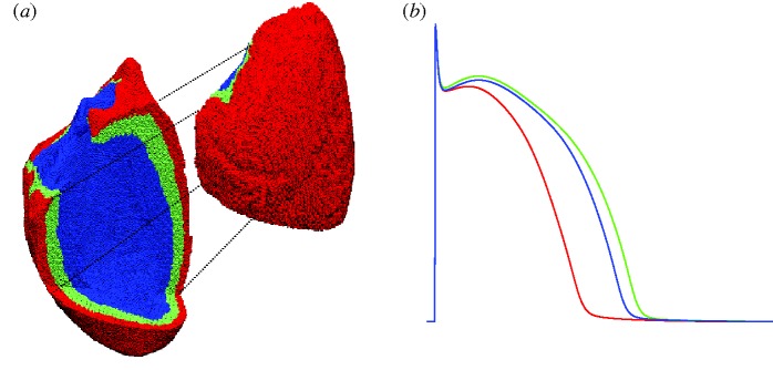
(a) The whole ventricular mesh. A portion of it is shifted to allow visualization of the three layers in which the wall has been subdivided: endocardial (blue), mid-myocardial (green) and epicardial (red). (b) Action potentials of isolated cells included in the three layers.