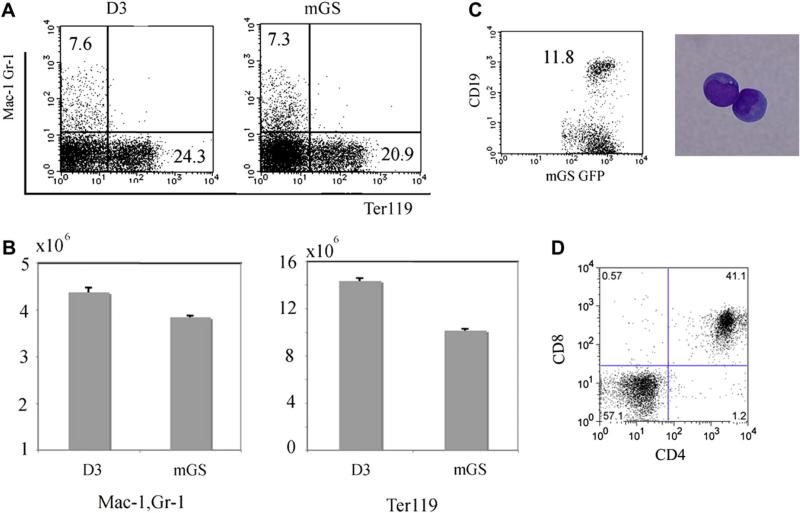
Figure 4.
Myelolymphoid potential of mGS cells. Mac1+Gr1+, Ter119+, CD19+, and CD4+CD8+ cells were differentiated from mGS-derived Flk-1+ cells within OP9 culture (A,B,C) or OP9-DL1 culture (D). For Mac1+Gr1+, Ter119+ cells, cells were collected from 8–10 days culture (A,B). The numbers of myeloid and erythroid cells differentiated from mGS and ES cells were similar (B). For CD19+ (C), and CD4+CD8+ cells (D), cells were collected from 14–21 days culture. These FACS data are representative among three independent experiments.

