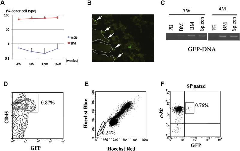
Figure 6.
Transplanted hematopoietic cells from GFP+ mGS cells can be detected in bone marrow (BM) 4 months after transplantation and displays stem cell phenotype. After transplantation, peripheral blood (PB) was analyzed every 4 weeks (A). BM cells from recipient mice 4 months after transplantation were analyzed by LSR II (D, E, F). GFP+CD45+ cells were detected (D). When the BM cells of the recipient mice were stained with Hoechst 33324, GFP+ cells were detected in the SP region (E, F). In the section of recipient BM, GFP+ cells were found attached to the endosteal region (B, arrow). RT-PCR shows donor-derived DNA in the BM and spleen at 7 weeks and 4 months after transplantation (C).

