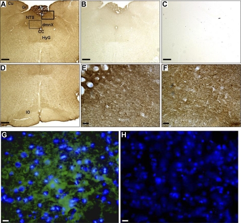
Fig. 6.
Immunolocalization of ANG-(1–12). Paraformaldehyde-fixed, frozen brain sections (n = 3 and 30 μm) were incubated with the ANG-(1–12) antibody to determine expression of ANG-(1–12) in dorsal medulla of Sprague-Dawley rats. 3,3′-Diaminobenzidine (A–F) and immunofluorescence (G and H) staining show ANG-(1–12) expression in various brainstem regions, including the NTS, in representative adjacent sections at ∼−13.8 mm caudal to bregma. ANG-(1–12) immunostaining was widely expressed in the medulla (A and D; 5×). Control sections (5×) were incubated with 40 μM ANG-(1–12) peptide together with the ANG-(1–12) antibody (B) or no ANG-(1–12) antibody (C). A higher magnification (40×) shows ANG-(1–12)-like immunoreactivity in cell bodies and fibers in the area postrema (E), NTS (E), and dorsal motor nucleus of the vagus (F). Adjacent sections were incubated with the ANG-(1–12) antibody (G) or no primary antibody (H) for immunofluorescence localization. ANG-(1–12) fluorescence is shown in green and nuclei in blue (40×). dmnX, Dorsal motor nucleus of the vagus; Cu, cuneate nucleus; Gr, nucleus gracilis; CC, central canal; HyG, hypoglossal nucleus; IO, inferior olivary nucleus. Scale bars: A–D, 100 μm, and E–H, 10 μm.

