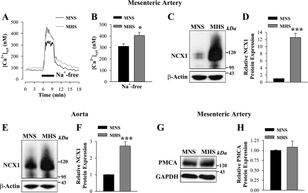
Fig. 5.
Enhanced Ca2+ entry via the reverse mode of Na+/Ca2+ exchanger-1 (NCX1) and augmented NCX1 expression in freshly dissociated myocytes from MHS rats. A and B: activation of the reverse mode of NCX1 in ASMCs from MNS (black) and MHS (gray) rats. A: representative time course records showing changes in [Ca2+]cyt in single ASMCs; time of treatment with Na+-free solution is indicated. Nifedipine (10 μM) was added 10 min before the records shown and was maintained throughout the experiment. B: summarized data show the NCX1-mediated Ca2+ entry in 33 MNS and 23 MHS mesenteric ASMCs. *P < 0.05 vs. MNS arterial myocytes. C–H: Western blot analysis of NCX1 (C–F) and plasma membrane Ca2+-ATPase (PMCA; G and H) protein expression (30 μg/lane) in smooth muscle cell membranes from mesenteric arteries (C, D, G, and H) and aortas (E and F) of MNS and MHS rats. C, E, and G: representative blots. Summary data (D, F, and H) are normalized to the amount of β-actin and are expressed as means ± SE from 9 (D), 18 (F), and 5 (H) immunoblots (total of 22 rats). ***P < 0.001 vs. MNS ASMCs.

