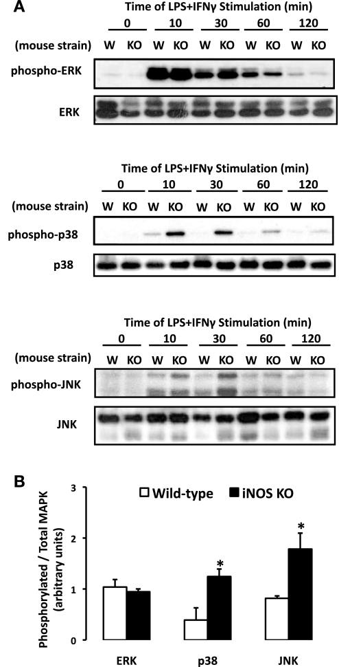
Fig. 6.
MAPK phosphorylation profile elicited by LPS + IFN-γ is modified in SMCs isolated from iNOS−/− vs. WT mice. A: SMCs were exposed to LPS + IFN-γ for 0–120 min, lysed, and probed for the basal and phosphorylated isoforms of ERK, p38, and JNK by Western blot analysis. W, WT; KO, knockout. B: MAPK phosphorylation (30 min) was quantified by densitometric analysis and expressed as phosphorylated-to-total MAPK ratio (means ± SE; *P ≤ 0.05 vs. WT).

