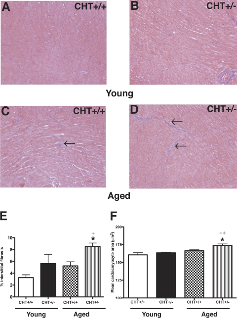
Fig. 7.
Cardiac interstitial fibrosis and mean myocyte alterations in CHT+/+ and CHT+/− mice. A–D: Masson's trichrome-stained 5-μm sections of the paraffin-embedded left ventricular myocardium from young (2–3 mo; A and B) and aged (10–12 mo; C and D) CHT+/+ mice (A and C) and CHT+/− mice (B and D). E: morphometrical analysis of myocyte fibrosis showed that aged CHT+/− mice exhibited significantly higher left ventricular fibrosis compared with aged CHT+/+ mice. *P < 0.01 and +P = 0.04, young vs. aged CHT+/− mice. F: morphometrical analysis of mean cardiomyocyte area from the periodic acid-Schiff-stained left ventricular myocardium showed similar cardiomyocyte area in young CHT+/+ and CHT+/− mice. However, aged CHT+/− mice displayed significantly increased cardiomyocyte area versus aged CHT+/+ mice. *P = 0.01 and ++P = 0.002, young vs. aged CHT+/− mice. Arrows reflect areas of cardiac interstitial fibrosis. Values are means ± SE; n = 5 mice/genotype. Significance was determined using a one-tailed, unpaired Student's t-test (genotype × same age group).

