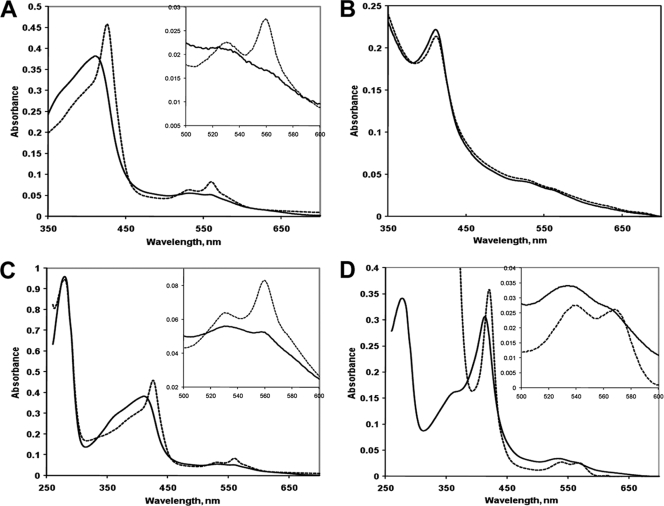
FIG. 2.
(A) Electronic absorption spectra of the MBP fusion to the SCHIC domain of the R. sphaeroides PpaA protein, MBP-SCHICRs, as purified from E. coli. Solid trace, original spectrum; dashed trace, spectrum obtained after addition of dithionite. (B) Spectra of MBP-SCHICRs purified from anaerobic E. coli cells grown in the absence (solid trace) and presence (dotted trace) of 50 μM cyanocobalamin. (C) Spectra of MBP-SCHICRs after reconstitution with hemin in vitro. Solid trace, original spectrum; dashed trace, spectrum obtained after reduction with dithionite (and dithionite removal by size exclusion chromatography). (D) Spectra of MBP-SCHICJs after reconstitution with hemin in vitro (approximately 1:1 molar ratio). Solid trace, original spectrum; dashed trace, spectrum obtained after reduction with dithionite. Insets show magnified long-wavelength regions.

