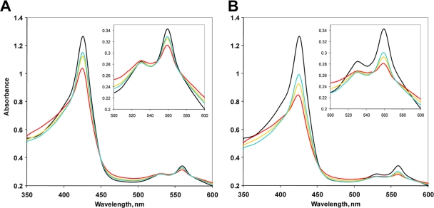
FIG. 3.
Electronic absorption spectra of MBP-SCHICRs before and after exposure to 100 μM oxygen (final concentration) (A) and 200 μM oxygen (B). The medium contained 5 mM dithiothreitol and a glucose-glucose oxidase-catalase system of oxygen removal (3). Black traces, deoxy, Fe2+ protein; red traces, protein immediately after addition of oxygen; gold traces, protein after 3 min; cyan traces, protein after 15 min. Insets show magnified long-wavelength regions. The experimental details are given in reference 23.

