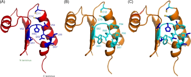
FIG. 3.
Homology structural model of S. aureus NrdH. (A and B) S. aureus NrdH model structure (see Materials and Methods) (A) and E. coli NrdH X-ray structure (PDB accession number 1H75) (B) (40) presented in cartoons. The two cysteines of the CXXC redox-active site are shown as sticks and are labeled. The residues of the loop connecting β4 to α3 are shown as sticks and are colored blue (A) and cyan (B). (C) Superposition of the two structures performed by using Multiprot (38). The RMSD between the two structures is 0.47 Å.

