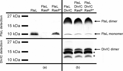
FIG. 1.
Coexpression of ftsL, divIC, yluC, and yluC(E21A) genes. Shown are immunoblots of 10% Schägger SDS system gels. For FtsL detection, blots were treated with anti-PentaHis and anti-mouse IgG alkaline phosphatase (AP), and for DivIC detection, they were treated with anti-DivIC and anti-rabbit IgG AP. Proteins present in each strain after expression are noted on top. (a) Influence of active RasP and inactive RasP-E21A (RasP*) on FtsL levels. (b) Influence of DivIC on FtsL oligomerization and stability. Asterisks indicate DivIC degradation bands.

