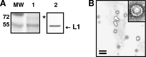
FIG. 2.
Characterization of purified HPV-16-SEAP PsV. (A) An aliquot of purified PsV preparation was analyzed by SDS-PAGE with Coomassie brilliant blue staining (lane 1) or immunoblotting with an anti-L1 antibody (B0580; Dako Corporation, Carpinteria, CA) (lane 2). The asterisk indicates a faint band that is probably the minor capsid protein L2. (B) Electron micrograph of a purified PV preparation. Bar, 100 nm. The inset shows a negatively stained PsV capsid at a higher magnification.

