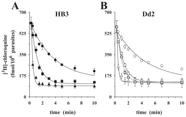
Fig. 4.
Chloroquine efflux kinetics under ‘reverse varying-trans’ conditions, in glucose-free medium. P. falciparum-infected erythrocytes were pre-loaded with comparable amounts of [3H]-chloroquine, washed and placed in glucose-free medium (replacing glucose by an equal amount of 2-deoxyglucose) containing the following concentrations of cold chloroquine: 0.0 μM (circles), 0.1 μM (squares), 1.0 μM (triangles).
A. CQS parasite HB3.
B. CQR parasite Dd2.
The grey squares (0.1 μM external chloroquine) show the effect of addition of 10 μM verapamil at zero time (dotted line). The means ± SEM of at least three independent determinations are shown.

