Fig. 4.
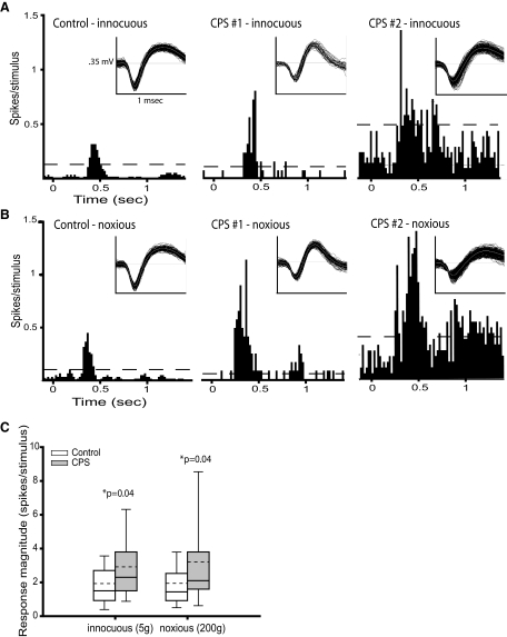
Neuronal activity in SI is enhanced in animals with CPS. A: peristimulus time histograms (PSTHs, 20 ms bins) showing the responses to innocuous mechanical hindpaw stimulation (5 g) of SI neurons from a sham-lesioned rat (left) and a spinal-lesioned rat with behaviorally confirmed CPS (middle and right). Sensory-evoked activity in SI neurons recorded from the CPS rat is markedly higher than in the neuron from the sham rat. Spontaneous activity is also higher in the second SI neuron from the CPS rat (right) than in the neuron from the sham rat. Dashed lines represent, the threshold at which the response significantly exceeded the spontaneous firing rate (99% confidence interval). Inset: representative spike waveforms. B: PSTHs showing the responses to noxious mechanical hindpaw stimulation (200 g) of SI neurons from a sham rat (left) and a CPS rat (middle and right). As in A, the sensory-evoked neuronal activity is higher in the CPS rats. C: group data showing that activity evoked by both innocuous and noxious mechanical stimulation of the hindpaw is significantly higher in SI neurons from animals with CPS (n = 30) than from control rats (n = 28). - - -, mean values.

