Fig. 6.
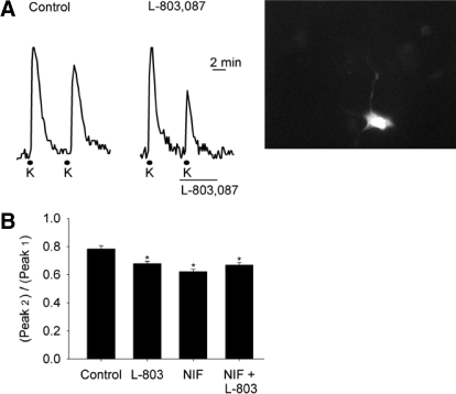
Intracellular Ca2+ signaling was reduced by L-803,087 in ganglion cells in retinal flat mount. A: applications of 52 mM K+ solution for 30 s depolarized ganglion cells and produced transient increases in intracellular Ca2+ concentration, measured with the dye fura-2. Traces (left) show that, in the absence of drug, the second of paired K+ applications produced a smaller peak Ca2+ signal. On the right, the paired K+ pulses, with L-803,087 (100 nM) applied 30 s prior to the second K+ application, show greater reduction of the Ca2+ signal compared with control. Far right panel shows a fluorescent image (stimulated at 380 nm) of a fura-2-containing ganglion cell in the flat-mount retina. B: mean data show amplitudes of the second high K+ response expressed as a percentage of first peak in control, L-803,087 (L-803, 100 nM), nifedipine (NIF, 10 μM), and nifedipine plus L-803,087. The mean Ca2+ transient amplitudes of all 3 drug treatment groups (L-803, NIF, L-803 + NIF) were significantly smaller than those of the control group, but they did not differ among themselves (n = 21–89 cells/group; *P < 0.05, ANOVA).

