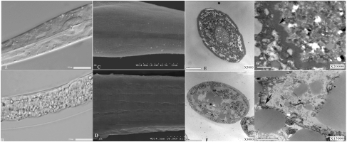
Fig. 2.
Microscopic examination of B. nematocida strain B16 target sites. (A) Both the intestine and cuticle of nematodes were intact when treated with E. coli. (B) Structures of pharynx, muscle, and intestine were disorganized when treated with B. nematocida strain B16. (C) Nematodes in the E. coli-treated control group had smooth undisturbed surfaces with a healthy cuticle structure that included the regular striae and lateral lines. (D) Nematodes infected with B. nematocida strain B16 showed a lightly exfoliated cuticle. (E) The cross-section of an untreated, healthy nematode showed a highly ordered and compact intestinal structure. (F) The cross-section of a nematode infected with B. nematocida strain B16 showed numerous defects including fusion, vesiculation, and loosening of various organs. (G) Low-magnification TEM of the midgut of the control nematode showed ordered, densely arrayed, and normal-looking microvilli. (H) Microvilli in strain B16-infected nematodes appeared destroyed with significant membrane-tethering defects. Arrows indicate healthy (G) and damaged (H) and microvilli.

