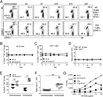
Fig. 2.
NP− B cells and CD4+ T cells maintain a 1:1 ratio. (A) Contribution of transferred B1-8 and B1-8.H2-O−/− cells to donor-derived NP− non-GC B-cell and CD4+ T-cell populations over time. Donor-derived cells were gated as shown in Fig. S1A. Numbers indicate percentage of each gated population. (B and C) Quantification of data shown in A for multiple mice. (D) Ratio of H2-O−/− to WT cells for populations shown in A, normalized to ratio present in transferred mixture. (E) B1-8 and B1-8.H2-O−/− B cells (1:1 mix) were transferred into recipient mice and either immunized with NP-CGG or not immunnized. Ten days later, the percentage of WT and H2-O−/− B1-8 cells were measured as shown in Fig. S2B and quantified for multiple mice. (F) Ratio of H2-O−/− to WT cells normalized to ratio of original mixture for data shown in E. (G) Purified and irradiated B cells from B1-8, B1-8.H2-O−/−, and nontransgenic (nTg) littermate mice were pulsed for 30 min on ice with increasing concentrations of NP-CGG, washed extensively, and cocultured for 72 h with NP-CGG–specific CD4 T cells. T cell proliferation was determined by [3H]thymidine incorporation during the last 18 h of culture. Data are representative of four independent experiments. Statistical significance determined by two-tailed paired t test for E and a two-tailed unpaired t test for F.

