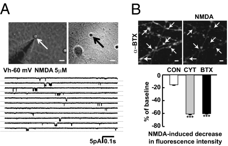
Fig. 6.
Presynaptic NMDAR function is enhanced in enlarged presynaptic boutons. (A) Examples of NMDA-activated channel currents recorded in outside-out membrane patches excised from enlarged presynaptic boutons in treated cultures. Similar channel currents were recorded upon application of 5 μM NMDA from five distinct terminals, and displayed a chord conductance of 58 ± 4 pS. (B) Fura-2 calcium imaging using two-photon microscopy at 780-nm excitation wavelength showed that NMDA stimulation led to calcium entry as indicated by decreased fluorescence intensity in presynaptic boutons of α-BTX–treated cultures. Quantification showed that NMDA stimulation produced a greater decrease in fluorescence intensity in the presence of tetrodotoxin and calcium channel blockers in boutons of α-BTX– (n = 48, P < 0.001 vs. control) or cytisine-treated (n = 44, P < 0.001 vs. control) cultures compared with those of control cultures (n = 12). (Scale bars, 5 μm.)

