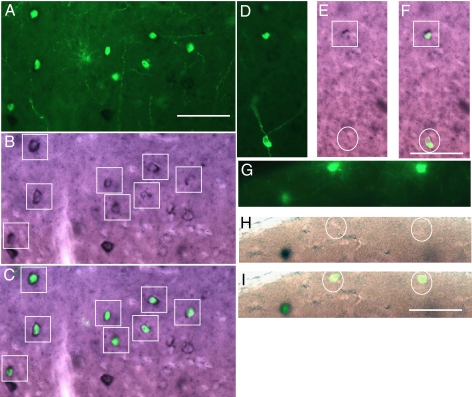
Fig. 2.
Double-labeling for GFP expression and ErbB4 in situ hybridization. GFP indicates cells infected with EnvB-pseudotyped virus and TVB–NRG1 bridge protein and stained with a heat-denatured anti-GFP antibody. Black reaction product indicates anti-digoxigenin staining after labeling with an ErbB4 antisense RNA probe. (A–C) Low power views indicating distribution of GFP-positive cells (A), ErbB4 in situ labeling (B), and overlay (C). Many of the GFP-positive cells are also ErbB4-positive. ErbB4-positive cells are marked by boxes (B, C, E, and F), and all of the GFP-positive cells can be seen to be double-labeled in the overlay (C). In some cases, GFP-expressing cells either did not have obvious ErbB4 label (D–F, circled cell) or were clearly not labeled (G–I, circled cells). (Scale bars: A, 50 μm and corresponds to A–C; F, 50 μm and corresponds to D–F; I, 50 μm and corresponds to G–I.)

