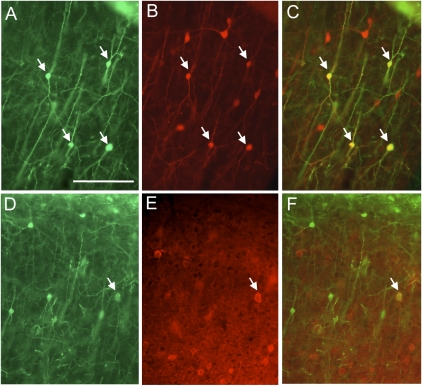
Fig. 4.
Double-labeling for GFP expression and inhibitory cell type-specific markers. Green label indicates anti-GFP staining of cells infected with EnvB-pseudotyped virus and TVB–NRG1 bridge protein, whereas red corresponds to antibody staining against CR (A–C) or PV (D–F). GFP expression in A and D illustrates overall location and morphologies of infected, GFP-labeled neurons. Pial surface is at the top. B and E correspond to the same sections to their left and illustrate red labeling with anti-CR (B) or PV (E). C and F are overlays of the GFP and antibody-stained images. Some of the double-labeled cells are marked by arrows. Note that GFP-expressing cells colabeled for CR are common, whereas labeling with PV is rare (selected photograph highlights on such cell). (Scale bar: A, 100 μm and corresponds to all images.)

