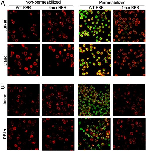
Fig. 4.
There is an internal pool of RBR receptor in suspension lymphocytes. (A) Jurkat T and Daudi B lymphocytes, which grow in suspension, were fixed, their plasma membranes were labeled with DiD (red) and either left intact (nonpermeabilized) or permeabilized with saponin, and then were incubated with Fc-conjugated RBR (500 nM) and anti-rabbit Fab 488 (green). Images were captured and processed as in Fig.1. (B) Human primary blood lymphocytes (PBLs) and Jurkat cells were processed as in A except that the γ level of the green channel was increased to 1.3 (for all images in B) to compensate for lower laser intensity settings on the microscope (percent laser power and gain). Findings represent results from two or more experiments examining at least five fields per sample.

