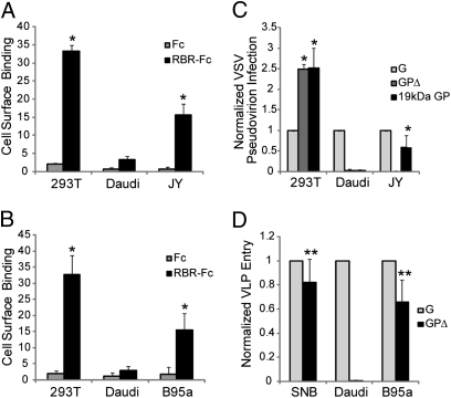
Fig. 5.
Adherent B lymphocytes bind RBR at the cell surface and are susceptible to EBOV-GP mediated entry and infection. (A) Adherent 293T and JY cells were lifted by pipetting with cold PBS++, whereas suspension Daudi cells were collected and chilled to 4 °C. Cells were then incubated with Fc-conjugated RBR (200 nM) followed by anti-rabbit Fab 488. Rabbit Fc was used as a negative control. Cell-surface binding was analyzed by flow cytometry. (B) 293T, Daudi, and B95a cells were examined for RBR cell-surface binding as in A. Data bars in A and B represent the average percent of cells showing RBR binding at their surface from three independent experiments (each with duplicate samples). Error bars represent SD. Statistical significance compared with RBR binding to the surface of Daudi cells was determined by Student t test (*P < 0.015). (C) 293T, Daudi, and JY cells were infected with VSV pseudovirions bearing either VSV G, EBOV GPΔ, or EBOV 19-kDa GP (6, 16). The average percent of cells infected with pseudovirions bearing VSV G, EBOV GPΔ, and EBOV 19-kDa GP were, respectively: 293T: 21%, 51%, and 53%; Daudi: 12%, 0%, and 0%; JY: 19%, 0%, and 10%. B95a cells are resistant to WT VSV infection (Fig. S5C) and thus, could not be examined in this experiment. (D) SNB19 (SNB), Daudi, and B95a cells were spinoculated with VLPs bearing VSV G or EBOV GPΔ as described in Materials and Methods. The average percent of cells showing entry with VLPs bearing VSV G and EBOV GPΔ were, respectively: SNB19: 91% and 75%; Daudi: 33% and 0%; B95a: 24% and 15%. JY cells could not be examined in this experiment because of high levels of background signal (Fig. S5D). In C and D, bars represent the average percent of cells infected (C) or showing entry (D) normalized to infection or entry by particles bearing VSV G in the same cell type from at least three experiments each with duplicate samples. Error bars represent SD of the normalized data. Statistical significance compared with infection or entry in Daudi cells was determined by Student t test (*P < 0.03; **P < 0.0002).

