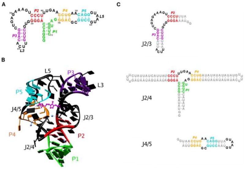
Figure 1.

The aptamer domain of the thiamine pyrophosphate riboswitch from the thiC gene in A. thaliana examined in this study. (A) Secondary structure diagram of the riboswitch construct. The five base-paired helices are labeled P1 – P5 and are each given a unique color: P1 (green), P2 (red), P3 (violet), P4 (yellow), P5 (cyan). The non base-paired regions – junctions and loops – are shown in black. (B) Structure of the TPP riboswitch aptamer as characterized previously by X-ray crystallography. The color scheme is as defined in (A). The ligand TPP is shown in magenta in a ball-and-stick representation. Mg2+ ions found in the crystal structure are represented as blue spheres. (C) Constructs of the three junctions in the structure of the aptamer. The junctions are defined by the adjoining helices: J2/3 between P2 and P3 (left), J2/4 between P1, P2 and P4 (middle), and J4/5 between P4 and P5.
