Figure 2.

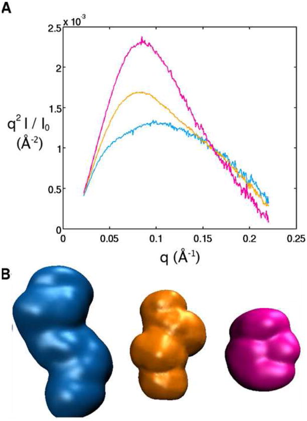
Small Angle X-ray Scattering (SAXS) data for the structure of the TPP riboswitch aptamer in the absence of Mg 2+ and TPP (blue) defined as the ‘unfolded’ state, in 10 mM Mg2+ but no TPP (orange) defined as the ‘intermediate’ state, and in 10 mM Mg2+ and 10 mM TPP (pink) defined as the ‘folded’ state. Data is also shown for the denatured riboswitch in 7 M urea (grey). (A) SAXS profiles in Kratky representation (q2I(q) vs. q for the full scattering profile) in the different solution conditions. (B) Low resolution bead models for the TPP riboswitch in its unfolded, intermediate and folded state, obtained from the SAXS profiles in (A). See Materials and Methods for details on the structure reconstruction.
