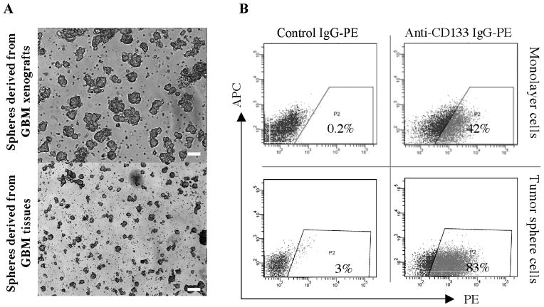
Fig. 1.
Characterization of GBM tumor spheres. A, Morphology of GBM tumor spheres derived from xenografts and fresh human tissues after 14 days of incubation in serum free restrictive media. Scale bar, 500 μm. B, CD133 epitope expression by GBM cells grown as a monolayer in serum containing media or as tumor spheres in serum free media.

