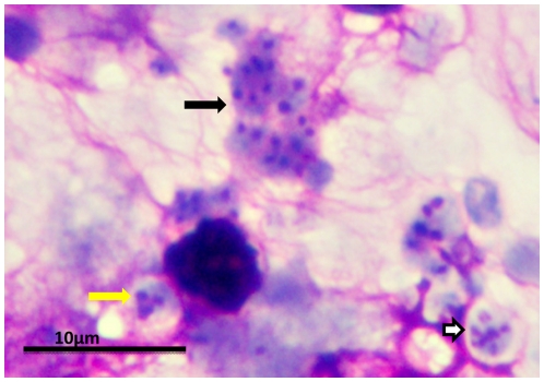
Figure 2. P. murina stained with rapid Wright-Giemsa.
Clusters of P. murina from a homogenate of an infected mouse lung were dropped on glass slides and stained with a rapid Wright-Giemsa. The black arrow points to a cluster of trophic forms. The white arrow indicates a mature cyst. The yellow arrow indicates an immature cyst with only three nuclei present in this section. The magnification bar represents 10 um. The micrograph was taken with an Olympus BH2 microscope and DP-72 digital camera.

