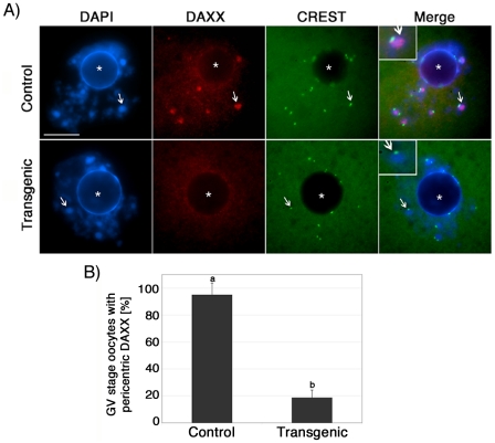
Figure 4. ATRX is required to recruit the transcriptional regulator (DAXX) to pericentric heterochromatin in mammalian oocytes.
(A) Top Panel: Nucleus of a control oocyte showing a precise co-localization of DAXX (red) with bright, DAPI-stained pericentric heterochromatin domains (arrows). The position of the centromere is indicated by CREST (green). Lower Panel: Analysis of transgenic oocytes demonstrated that in the absence of ATRX, the transcriptional regulator DAXX fails to associate with pericentric heterochromatin domains while nucleoplasmic expression of DAXX persists. The position of the nucleolus is indicated by (*). (B) Proportion of germinal vesicle (GV) stage oocytes showing pericentric DAXX localization. More than 80% of transgenic oocytes fail to recruit DAXX to pericentric heterochromatin. Data are presented as the mean ± s.d. of three independent experiments. CREST immunolocalization (green) was conducted as an experimental control for centromeric-kinetochore integrity. Scale bars = 10 µm.

