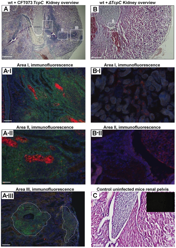
Figure 2. Deletion of TcpC abrogates abscess formation in kidney sections from wild type mice infected with CFT073.
(A) Overview of htx-eosin stained kidney section of wt mice infected with CFT073, showing abscesses (scale bar, 500 µm). Magnified areas of section A shown as A-I, A-II and A-III are stained with specific antibodies to neutrophils (green, NIMP-R14) and the PapG adhesin (red, antiserum to a synthetic PapG peptide) (scale bar, 100 µm). Abscesses in wt mice are shown by dotted lines. (B) Overview of kidney section of wt mice infected with ΔTcpC (scale bar, 300 µm). Magnified areas of section B shown as areas B-I and B-II are stained with specific antibodies as described above. (C) Kidney section of uninfected control mice with htx-eosin staining (scale bar, 200 µm) and inset picture showing negative control for anti-neutrophil and anti-PapG antibodies.

