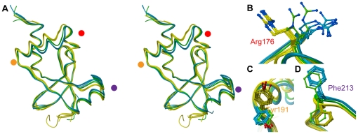
Figure 4. Conformational changes among the SnoN-DHD monomers.
Panel A: Superposition of the twelve monomers of SnoN-DHD present in the asymmetric unit (stereo view). Chains A, B, C, D, E and G are colored in yellow, chains F, I, K and L in blue, and chains H and J in green. The red, orange and violet dots represent the location of Arg176, Tyr191 and Phe213, respectively. Panels B–D: Side-chain conformational changes of Arg176, Tyr191 and Phe213.

