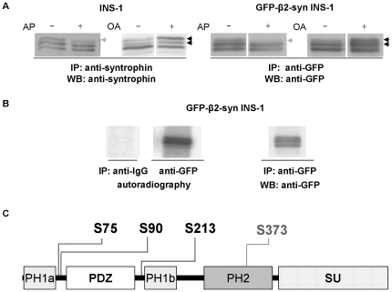
Figure 4. Phosphorylation/dephosporylation of β2-syntrophin.
A) Immunoblots with the anti-syntrophin or anti-GFP antibodies following immunoprecipitations with the same antibodies from extracts of INS-1 and GFP-β2-syntrophin INS-1 cells, respectively. Alkaline phosphatase (AP) was added to the immunoprecipitates, while okadaic acid (OA) was added before cell extraction. β2-syntrophin species the levels of which are reduced upon AP treatment are marked with a gray arrow, while black arrows indicated species increased upon OA incubation. B) The two panels on the left show the autoradiographies of 32P-GFP-β2-syntrophin immunoprecipitated with the rabbit anti-GFP antibody from extracts of GFP-β2-syntrophin INS-1 cells labeled with 32P kept in culture media. A control immunoprecipitation from the same cells was performed using rabbit control IgG. The right panel shows the immunoblot with the anti-GFP antibody on the same immunoprecipitated material visualized by autoradiography. C) Domain structure of β2-syntrophin, including the phosphoserines identified by mass spectrometry. PH = Pleckstrin Homology domain, PDZ = PSD95/Dlg/ZO-1 domain, SU = Syntrophin Unique domain.

