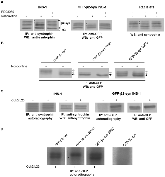
Figure 7. Phosphorylation of endogenous and GFP-β2-syntrophin and the S/D mutants by Cdk5.
A) Immunoblots with anti-syntrophin or anti-GFP antibodies on rat islet extracts (right panel) or immunoprecipitates obtained with the same antibodies from extracts of INS-1 cells (left panel) or GFP-β2-syntrophin INS-1 cells (middle panel) treated with the Cdk5 inhibitor roscovitine or the Erk1/2 inhibitor PD98059. Asterisks indicate the β2-syntrophin species sensitive to roscovitine. B) Immunoblots with the anti-GFP antibody on immunoprecipitates obtained with the same antibody from extracts of INS-1 cells expressing GFP-β2-syntrophin variants and treated with roscovitine. Gray and black arrows point to β2-syntrophin species the levels of which decreased or increased in response to roscovitine, respectively. C) Autoradiographies and immunoblots of β2-syntrophin (left panels) and GFP-β2-syntrophin (right panels) immunoprecipitated with anti-syntrophin or anti-GFP antibodies from INS-1 and GFP-β2-syntrophin INS-1 cells, respectively. Immunoprecipitates were incubated with or without the Cdk5/p25 complex in the presence of 32P-γ-ATP. D) Autoradiography of GFP-β2-syntrophin variants immunoprecipitated with the anti-GFP antibody from GFP-β2-syntrophin INS-1 cells and incubated with the Cdk5/p25 complex as in C. An asterisk indicates the β2-syntrophin species that is lacking in the GFP-β2-syntrophin S75D mutant.

