
Figure 1.
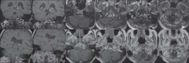
Row 1: At the time of the gamma knife radiosurgery (GKRS), the patient had a meningioma with a volume of 6.8 cc extending en plaque from the junction of the medulla and the pons into the foramen magnum along the clivus. Row 2: On follow-up imaging 13 years after GKRS, the lesion decreased in size to 4.0 cc without any new growth or radiographic complications of GKRS (cyst formation, necrosis, and edema)