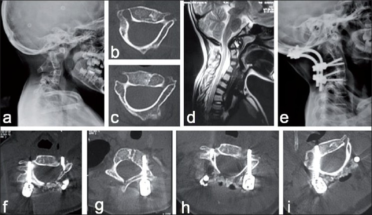
Figure 4.

Cervical pedicle screw-instrumented occipito-cervical fusion in an eight-year-old child. (a): Lateral radiograph of the cervical spine showing cervical segmentation anomaly with atlanto-axial instability. (b and c): Axial CT images showing atypical attenuated dysmorphic pedicles at C3 and C4 levels (d): Sagittal MRI of the cervical spine showing atlanto-axial instability with cord compression at the craniovertebral junction. (e): Lateral X-ray of the cervical spine shows reduction of atlanto-axial instability with subaxial cervical pedicle screws and occipito-cervical fusion. (f-i): Axial CT images showing well-contained cervical pedicle screws without any pedicle breach at C3, C4 levels
