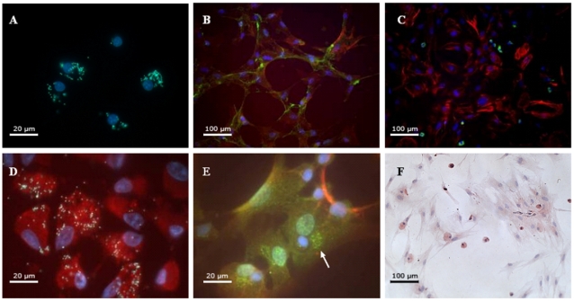
Figure 5. Isolation and differentiation of CD68+ cells.
(A) Cellular phagocytosis of yellow/green fluorescent particles by the purified CD68+ cell population from FBR. (B) ASMA (red) and smoothelin (green) expression in FBR CD68+ cells cultured for 21 days in complete Mesencult medium. (C) ASMA (red) and CD68 (green) expression of isolated cells cultured for 21 days in complete Mesencult medium. Small CD68+ cells and larger ASMA+ cells can be observed. (D) Functional macrophages that have engulfed yellow fluorescent particles show an expression of ASMA (red) as well as a morphological myofibroblast aspect. (E) Double staining for ASMA (red) and CD34 (green). The presence of intracytoplasmic CD34 expression can be clearly seen (white arrow). (F) Picture showing TGF-β receptor type II staining of a 42 days old CD68+ cell culture. TGF-β receptor type II staining appears in red-brown and nuclei appear in blue. The objective lenses used are Plan-APOCHROMAT 100×/1.4 (A, D, E) and Plan-APOCHROMAT 20×/1.4 (B, C, F).

