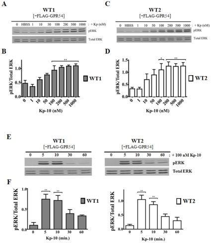
Figure 1. GPR54 is coupled to ERK MAPK pathway in WT MEFs.
Representative autoradiographs (A and C) and densitometric analyses (B and D) showing the expression of total and activated ERK1/2 in GPR54 overexpressing WT1 (β-arrestin-1 KO parent) and WT2 (β-arrestin-2 and 1/2 KO parent) MEFs following 10-minute treatment with increasing concentrations of Kp-10 (0–1000 nM). Representative autoradiograph (E) and densitometric analysis (F) showing the expression of total and activated ERK1/2 in GPR54 overexpressing WT1 and WT2 parental MEF cell lines following 100 nM Kp-10 treatment (for the indicated time points: 0, 5, 10, 30 and 60 minutes). Western blot analyses were done using monoclonal anti-ERK1/2 and anti-phospho ERK1/2 antibodies. The data represent the mean ± S. E. of 4 independent experiments. *P<0.05; **P<0.01vs control (0 min.).

