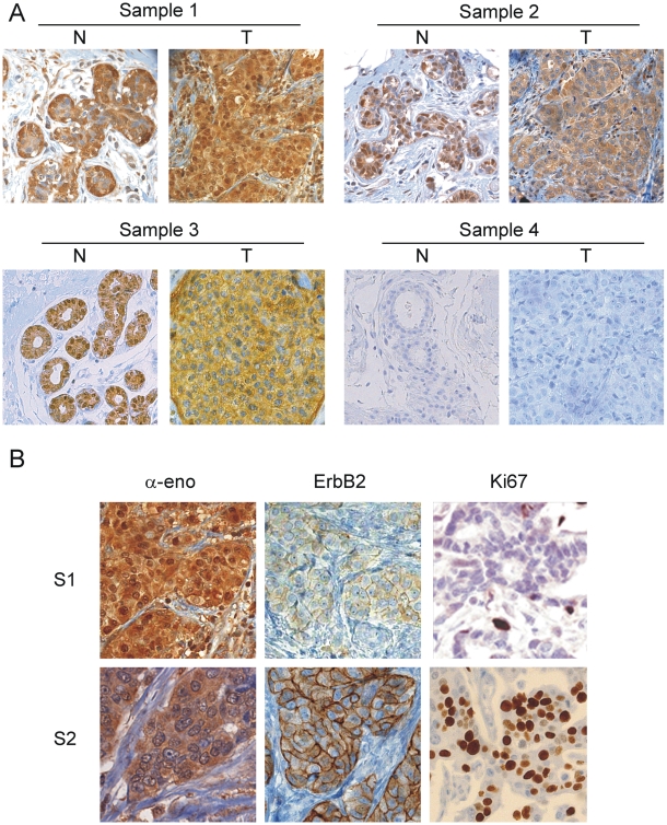
Figure 3. IDC immunohistochemical staining for α-enolase and nuclear MBP-1 revealed heterogeneity in breast cancer samples.
A. Representative immunohistochemical staining of 3 out of the 177 IDC samples analyzed with mAbs ENO-19/8. Tumor sections (T) were compared with the corresponding normal breast tissue (N). All normal breast tissues showed nuclear MBP-1 expression and low expression of cytoplasmic α-enolase (sample 1–3). In sample 1 and 2, cytoplasmic staining of the invasive breast carcinoma (T) was stronger than in the matched normal breast tissues. In tumor samples 2 and 3, the loss of MBP-1 nuclear expression occurred. Sections of sample 4 were immunoassayed with antibodies blocked with a recombinant α-enolase polypeptide (see materials and methods). Magnification: 250×. B. MBP-1 nuclear staining correlated with ErbB2 and Ki67 expression. Immunohistochemical staining for α-enolase, ErbB2 and Ki67 in MBP-1-positive (S1) and MBP-1-negative (S2) tumors. Magnification: 500×.

