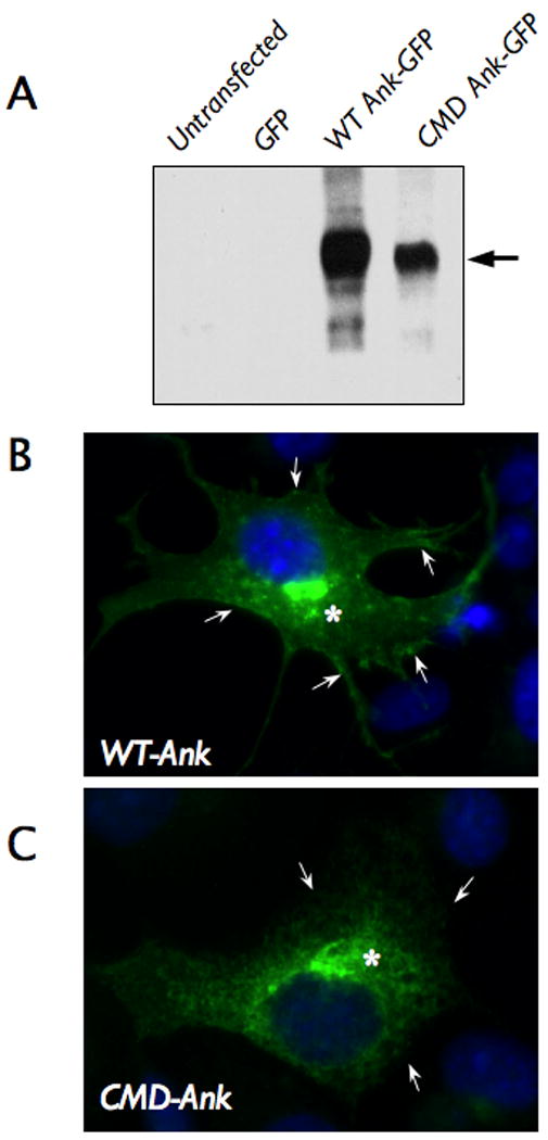
Figure 3.

Expression of Ank protein containing the craniometaphyseal dysplasia (CMD) mutation. A. Immunoblot analysis of COS-7 cells transfected with a mammalian expression vector encoding wild type murine Ank and green fluorescent protein (GFP) (WT Ank-GFP), or Ank containing the CMD mutation and GFP (CMD Ank-GFP). Untransfected cells and the cells transfected with a vector expressing GFP alone were used as controls. B&C. Immunofluorescent labeling of cells transfected with WT-Ank or CMD-Ank construct demonstrated that WT-Ank was localized in both plasma membrane (→) and cytoplasm (*) (B), while CMD-Ank was detected only in the cytoplasmic compartment as indicated with arrows (C).
