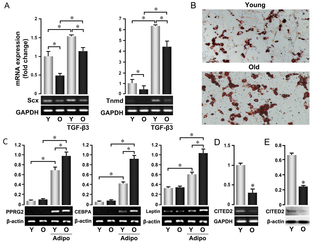
Figure 2. Altered cell fate of aged TSPCs.
P0 TSPCs were prepared as described in Figure 1 and used for all assays. (A) Expression of tendon lineage-specific genes. TSPCs were cultured with or without TGF-β3 for 3 days, and total RNA was extracted after culture. (B, C) Adipocyte-skewed differentiation of aged TSPCs. Cells were cultured in specific induction medium for 16 days. B: Oil Red O staining per (Gimble et al. 1995); C: mRNA expression of adipogenic marker genes in untreated and adipogenic-induced TSPCs. (D, E) Expression of Cited2 at mRNA (D) and protein (E) levels in young and old TSPCs. mRNA expression of indicated genes in A, C, D was assessed by RT-PCR. Upper Panels represent the quantification of band intensity from the gel shown in lower panels. Data are representative of 3 experiments and confirmed by real-time PCR.

