
Fig. 3.
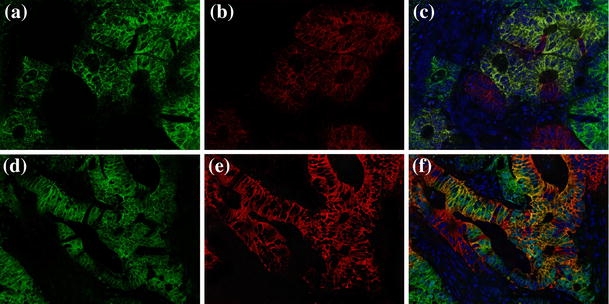
Double-label immunofluorescence staining for HIF-1α and P-gp in human colon carcinoma tissues. a–c, (×200), d–f, (×400). (green shows HIF-1α, red shows P-gp, yellow shows coexpression)
Official websites use .gov
A
.gov website belongs to an official
government organization in the United States.
Secure .gov websites use HTTPS
A lock (
) or https:// means you've safely
connected to the .gov website. Share sensitive
information only on official, secure websites.
Double-label immunofluorescence staining for HIF-1α and P-gp in human colon carcinoma tissues. a–c, (×200), d–f, (×400). (green shows HIF-1α, red shows P-gp, yellow shows coexpression)