
Figure 3.
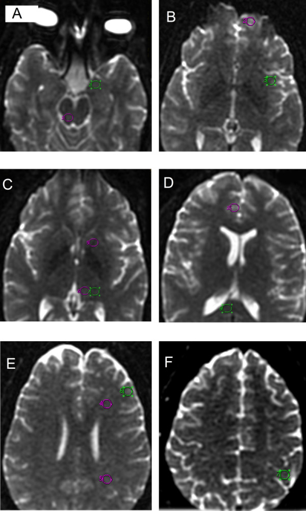
Axial images showing the different locations of the equal-sized regions of interest (ROIs). The ROI placements for the periaqueductal gray and amygdale (a), the orbitofrontal cortex and insular cortex (b), the internal capsule and ventral and dorsolateral thalamus (c), the gyrus cortex and corpus callosum (d), the frontal white matter, parietal white matter, and dorsolateral prefrontal cortex (e), and the sensorimotor area (f).