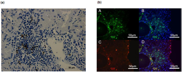
Figure 4.

Kidney CD134+ CD3+ infiltrating T-cells in lupus nephritis. (a) Representative renal biopsy of an SLE patient with lupus nephritis (WHO class IV). The biopsy was stained for CD134 using immunohistochemistry. (b) Staining of the biopsy for CD3+ and CD134+ cells by immunofluorescence. CD3 (A), CD3/DAPI (B), CD134 (C) and colocalization of CD134 with CD3 (D).
