
Figure 2.
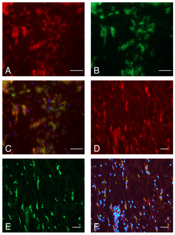
Expression of CCR9 by macrophages in rheumatoid synovium. Cryosections were treated with antibodies to CCR9 and CD14 or CD68 as markers of macrophages and double label immunofluorescence microscopy performed. (a) Shows staining for CCR9 (red) and (b) shows the same area stained for CD14 (green). (c) Shows a merge of images (a) and (b) and cells with colocalised CCR9 and CD14 (yellow). (d, e) Demonstrate staining for CCR9 and CD68 respectively and (f) is a merge of these two images, cells in yellow express CCR9 and CD68. Cell nuclei stain (blue) with DAPI in (c) and (f). The bar represents 50 μm.