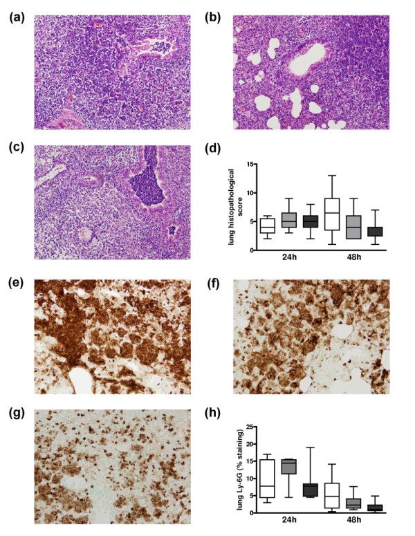
Figure 2.

Lung histopathology and neutrophil influx in untreated pneumococcal pneumonia. Lung haematoxylin and eosin staining 48 hours after pneumococcal pneumonia in (a) wild-type mice, (b) mice heterozygous and (c) mice homozygous for the factor V Leiden mutation (original magnification × 100). (d) Total lung pathology score 24 and 48 hours after induction of pneumococcal pneumonia in wild-type mice (white white, n = 8) and mice heterozygous (light grey white, n = 8) or homozygous (dark grey white, n = 8) for the factor V Leiden mutation. Representative slides of lung Ly-6G staining (brown) 48 hours after induction of pneumococcal pneumonia in (e) wild-type mice, (f) mice heterozygous and (g) mice homozygous for the factor V Leiden mutation (original magnification × 100). (h) Quantitation of pulmonary Ly-6G content 24 and 48 hours after induction of pneumococcal pneumonia in wild-type mice (white white, n = 8), and mice heterozygous (light grey white, n = 8) or homozygous (dark grey white, n = 8) for the factor V Leiden mutation. Data are expressed as box-and-whisker diagrams depicting the smallest observation, lower quartile, median, upper quartile and largest observation. There were no statistical differences between the groups at either time point.
