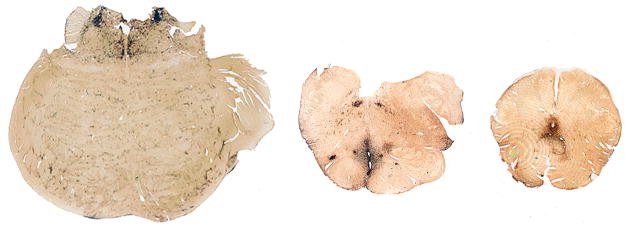
Figure 2.
(A–C) Whole mount 50-μm coronal sections of superior frontal cortex from case 1 (A), case 2 (B), case 3 (C) immunostained for tau with monoclonal antibody CP-13 showing extensive immunoreactivity that is greatest at sulcal depths (asterisks) and is associated with contraction of the cortical ribbon. (D–F) Microscopically there are dense tau-immunoreactive neurofibrillary tangles (NFTs) and neuropil neurites throughout the cortex, case 1 (D), case 2 (E) and case 3 (F). There are focal nests of NFTs and astrocytic tangles around small blood vessels (E, arrow) and plaque-like clusters of tau-immunoreactive astrocytic processes distributed throughout the cortical layers (F, arrows).

